

ART-LP02-04 · ART-LP02
Compare pelvic imaging methods by the structures and questions they can address without treating every image finding as a cause of infertility. Clear decisions begin by separating what is observed, why it matters, how the process works and which uncertainty remains.
Visual lesson summary
Review the lesson as a carousel.
Swipe or scroll through the key ideas, then continue with the detailed guidance below.
01 / 07Imaging the Uterus Ovaries and Pelvis 
02 / 07Why it matters 
03 / 07How the method works 
04 / 07How to read the evidence 
05 / 07What this cannot decide 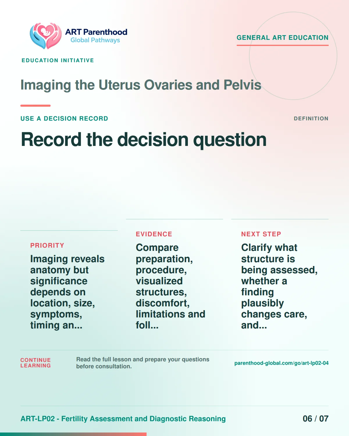
06 / 07Use a decision record 
07 / 07Keep the evidence visible
Define the exact question
transvaginal and transabdominal ultrasound, saline infusion sonography, selected MRI use, follicle and endometrial observations, fibroids, polyps, cysts and incidental findings.
Precision starts by defining the object, method and decision separately. For imaging the uterus ovaries and pelvis, useful records include ultrasound resolution, operator dependence, cycle-phase effects on endometrium, Doppler limitations. Each item should state who produced it, when it was produced, what population or specimen it represents, and which conclusion it can support. A familiar label may hide different assays, laboratory policies, legal meanings or endpoints, so the reader should ask for the operational definition rather than infer one from the name.
Why the distinction changes decisions
Imaging reveals anatomy but significance depends on location, size, symptoms, timing and treatment goals; incidental abnormalities can trigger unnecessary alarm or intervention.
The practical consequence is specific: misunderstanding imaging the uterus ovaries and pelvis can change which question is asked, which comparison appears favourable, or who seems to own the decision. Separate observed facts from interpretation and interpretation from choice. Record what remains unknown, what would change the conclusion and which excluded question belongs elsewhere: Tubal patency testing; Surgical treatment recommendations; Embryo-transfer ultrasound guidance. This keeps uncertainty visible without turning it into either alarm or reassurance.
How the process should work
Compare preparation, procedure, visualized structures, discomfort, limitations and follow-up, then show how clinicians reconcile images with history and other evidence.
Then test the method against one routine case and one discordant or incomplete case. Record where ultrasound resolution, operator dependence, cycle-phase effects on endometrium enter the sequence, who interprets them, what can delay the next step and which result would require the question to be reframed rather than forced into a yes-or-no answer.
Read measures without overreaching
Advanced interpretation should address ultrasound resolution and operator dependence, cycle-phase effects on endometrium, Doppler limitations, saline cavity contrast, lesion classification, interobserver agreement and incidentaloma bias.. The purpose is to show how the method works, where variation enters, which comparisons are defensible and what the evidence cannot establish. Keep ultrasound resolution, operator dependence, cycle-phase effects on endometrium, Doppler limitations, saline cavity contrast tied to their source, population and decision context; avoid universal thresholds, retrospective certainty and individual predictions from population averages.
Match evidence to the claim
Evidence must fit the exact claim in imaging the uterus ovaries and pelvis. Guidance can describe consensus or recommended process; a registry can describe observed outcomes; a systematic review can synthesize eligible studies; and a primary study can test a narrower question. Check version, population, endpoint, denominator, missing data, uncertainty and transferability before treating a source as decisive.
Trace each public statement to a stable claim ID and the source records that support it. Compare ultrasound resolution, operator dependence, cycle-phase effects on endometrium, Doppler limitations only when methods and populations are sufficiently alike. If a source addresses process but not effectiveness, safety but not legal effect, or a group average but not individual prediction, state that boundary directly.
Keep professional roles visible
For imaging the uterus ovaries and pelvis, professional roles are limited and complementary. An editorial reviewer checks scope discipline, plain-language accuracy, accessibility and whether wording overstates the evidence. A qualified clinician checks clinical terminology, interpretation limits, safety boundaries and escalation language. None of these roles replaces the informed choice of the person whose body, gametes, embryos, records, legal position or family life is affected. Record disagreements and conflicts of interest instead of hiding them behind a collective recommendation.
Build a decision record
Clarify what structure is being assessed, whether a finding plausibly changes care, and whether confirmation, surveillance, specialist review, or no action is appropriate.
A usable decision record for imaging the uterus ovaries and pelvis names the exact question, the affected person, the available options, the evidence and its limits, the professional responsible for interpretation, and the condition that would reopen the choice. It also records what is not yet known and whether the next step is reversible. The record should never convert a population estimate into a personal forecast, a laboratory category into a guarantee, a program policy into consent, or one jurisdiction's rule into universal law.
- Clarify what structure is being assessed, whether a finding plausibly changes care, and whether confirmation, surveillance, specialist review, or no action is appropriate.
- Confirm the source and update date for imaging, uterus, ovaries.
- Record what pelvis, explain, transvaginal can and cannot decide.
- Route unresolved questions to editorial, medical.
For Nerds: Technical Deep Dive
Cover ultrasound resolution and operator dependence, cycle-phase effects on endometrium, Doppler limitations, saline cavity contrast, lesion classification, interobserver agreement and incidentaloma bias.
Mechanism, measurement and endpoint
Cover ultrasound resolution and operator dependence, cycle-phase effects on endometrium, Doppler limitations, saline cavity contrast, lesion classification, interobserver agreement and incidentaloma bias. Advanced interpretation starts by defining construct, measurement and endpoint. The relevant technical vocabulary includes imaging, uterus, ovaries, pelvis, explain, transvaginal, transabdominal, ultrasound, saline, infusion, sonography, selected. These terms describe different layers: biological mechanism, observable signal, operational category, decision threshold and patient-relevant outcome. A strong analysis does not move between those layers without evidence. It records specimen or document provenance, analytical method, timing, comparison population, missingness, uncertainty and the professional who owns interpretation. It also asks whether the source is guidance, regulation, registry data, systematic review or primary research, because each supports different inferences. For selected, preserve the numerator, denominator, reference frame and failure modes. Test sensitivity, specificity, calibration, interobserver variation, selection bias, confounding and jurisdictional drift can each make a technically correct statement misleading in another context. A reviewer should verify current terminology and identify the evidence that would change the decision rather than adding unsupported precision.
- Explain transvaginal and transabdominal ultrasound, saline infusion sonography, selected MRI use, follicle and endometrial observations, fibroids, polyps, cysts and incidental findings.
- Compare preparation, procedure, visualized structures, discomfort, limitations and follow-up, then show how clinicians reconcile images with history and other evidence.
- Clarify what structure is being assessed, whether a finding plausibly changes care, and whether confirmation, surveillance, specialist review, or no action is appropriate.
Expected ranges / examples
- Topic-specific interpretation sequence: imaging -> uterus -> ovaries -> pelvis -> explain. A non-numeric process example showing why adjacent observations and decisions must not be treated as equivalent. Source: ASRM fertility evaluation.
Methods, categories and uncertainty
Compare preparation, procedure, visualized structures, discomfort, limitations and follow-up, then show how clinicians reconcile images with history and other evidence. Advanced interpretation starts by defining construct, measurement and endpoint. The relevant technical vocabulary includes imaging, uterus, ovaries, pelvis, explain, transvaginal, transabdominal, ultrasound, saline, infusion, sonography, selected. These terms describe different layers: biological mechanism, observable signal, operational category, decision threshold and patient-relevant outcome. A strong analysis does not move between those layers without evidence. It records specimen or document provenance, analytical method, timing, comparison population, missingness, uncertainty and the professional who owns interpretation. It also asks whether the source is guidance, regulation, registry data, systematic review or primary research, because each supports different inferences. For imaging, preserve the numerator, denominator, reference frame and failure modes. Test sensitivity, specificity, calibration, interobserver variation, selection bias, confounding and jurisdictional drift can each make a technically correct statement misleading in another context. A reviewer should verify current terminology and identify the evidence that would change the decision rather than adding unsupported precision.
- Explain transvaginal and transabdominal ultrasound, saline infusion sonography, selected MRI use, follicle and endometrial observations, fibroids, polyps, cysts and incidental findings.
- Compare preparation, procedure, visualized structures, discomfort, limitations and follow-up, then show how clinicians reconcile images with history and other evidence.
- Clarify what structure is being assessed, whether a finding plausibly changes care, and whether confirmation, surveillance, specialist review, or no action is appropriate.
Expected ranges / examples
- Topic-specific interpretation sequence: uterus -> ovaries -> pelvis -> explain -> transvaginal. A non-numeric process example showing why adjacent observations and decisions must not be treated as equivalent. Source: ASRM fertility evaluation.
Limits, review and decision ownership
Clarify what structure is being assessed, whether a finding plausibly changes care, and whether confirmation, surveillance, specialist review, or no action is appropriate. Advanced interpretation starts by defining construct, measurement and endpoint. The relevant technical vocabulary includes imaging, uterus, ovaries, pelvis, explain, transvaginal, transabdominal, ultrasound, saline, infusion, sonography, selected. These terms describe different layers: biological mechanism, observable signal, operational category, decision threshold and patient-relevant outcome. A strong analysis does not move between those layers without evidence. It records specimen or document provenance, analytical method, timing, comparison population, missingness, uncertainty and the professional who owns interpretation. It also asks whether the source is guidance, regulation, registry data, systematic review or primary research, because each supports different inferences. For explain, preserve the numerator, denominator, reference frame and failure modes. Test sensitivity, specificity, calibration, interobserver variation, selection bias, confounding and jurisdictional drift can each make a technically correct statement misleading in another context. A reviewer should verify current terminology and identify the evidence that would change the decision rather than adding unsupported precision.
- Explain transvaginal and transabdominal ultrasound, saline infusion sonography, selected MRI use, follicle and endometrial observations, fibroids, polyps, cysts and incidental findings.
- Compare preparation, procedure, visualized structures, discomfort, limitations and follow-up, then show how clinicians reconcile images with history and other evidence.
- Clarify what structure is being assessed, whether a finding plausibly changes care, and whether confirmation, surveillance, specialist review, or no action is appropriate.
Key takeaways
- transvaginal and transabdominal ultrasound, saline infusion sonography, selected MRI use, follicle and endometrial observations, fibroids, polyps, cysts and incidental findings.
- Imaging reveals anatomy but significance depends on location, size, symptoms, timing and treatment goals; incidental abnormalities can trigger unnecessary alarm or intervention.
- Compare preparation, procedure, visualized structures, discomfort, limitations and follow-up, then show how clinicians reconcile images with history and other evidence.
- Clarify what structure is being assessed, whether a finding plausibly changes care, and whether confirmation, surveillance, specialist review, or no action is appropriate.
FAQ
What exactly is Imaging the Uterus Ovaries and Pelvis?
transvaginal and transabdominal ultrasound, saline infusion sonography, selected MRI use, follicle and endometrial observations, fibroids, polyps, cysts and incidental findings.
Why does the distinction matter?
Imaging reveals anatomy but significance depends on location, size, symptoms, timing and treatment goals; incidental abnormalities can trigger unnecessary alarm or intervention.
How should the review work?
Compare preparation, procedure, visualized structures, discomfort, limitations and follow-up, then show how clinicians reconcile images with history and other evidence.
What belongs in the advanced evidence review?
ultrasound resolution and operator dependence, cycle-phase effects on endometrium, Doppler limitations, saline cavity contrast, lesion classification, interobserver agreement and incidentaloma bias.
What is outside this scope?
This package does not decide Tubal patency testing; Surgical treatment recommendations; Embryo-transfer ultrasound guidance. Those questions require their own evidence, scope and responsible professional.
What should be recorded before a decision?
Clarify what structure is being assessed, whether a finding plausibly changes care, and whether confirmation, surveillance, specialist review, or no action is appropriate.