
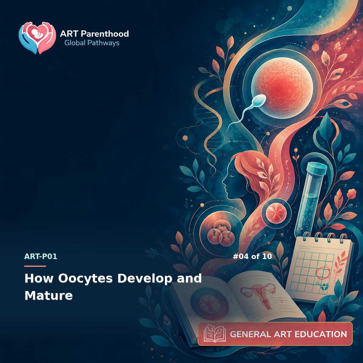
ART-LP01-04 · ART-LP01
Understand the oocyte life course and maturation stages so egg numbers, maturity, age effects, and laboratory reports are not mistaken for guaranteed competence. The useful starting point is to separate structures, processes, measurements and outcomes, then connect only the claims that biology and evidence can support.
Visual lesson summary
Review the lesson as a carousel.
Swipe or scroll through the key ideas, then continue with the detailed guidance below.
01 / 07How Oocytes Develop and Mature 
02 / 07Name each stage 
03 / 07Trace the mechanism 
04 / 07Read the measure 
05 / 07Keep the limit visible 
06 / 07Ask the next question 
07 / 07Use the full guide
Build the functional map
Oocyte development begins before birth, when germ cells enter meiosis and become enclosed in primordial follicles. Most remain arrested in prophase of meiosis I for years. The ovarian follicle pool declines through atresia as well as ovulation, and individual follicles spend months progressing from primordial to preantral and antral stages. The small antral cohort visible in one cycle is therefore not the whole reserve, and the reserve is not a count of future babies or usable embryos.
The ovarian reserve is a changing pool of follicles, while only a small cohort becomes visible and recruitable in a given cycle. Keep this observation tied to its collection time, method and biological stage; the next inference requires evidence that directly answers the clinical question. For every number, ask whether it refers to follicles, oocytes retrieved, mature MII oocytes, normally fertilized oocytes or embryos.
Follow the biological sequence
A follicle is a multicellular ovarian structure; an oocyte is the germ cell within it. Granulosa and theca cells support growth, steroid production and communication with the oocyte. As antral follicles become gonadotropin-responsive, one is usually selected in an unstimulated cycle, while treatment may support several. Ultrasound measures fluid-filled follicles rather than seeing the oocyte itself. Empty or small follicles, failed recovery and variable maturity explain why follicle counts and retrieved-oocyte counts need not match.
Oocytes remain arrested in meiosis for years and must coordinate nuclear and cytoplasmic maturation before fertilization. Keep this observation tied to its collection time, method and biological stage; the next inference requires evidence that directly answers the clinical question. For every number, ask whether it refers to follicles, oocytes retrieved, mature MII oocytes, normally fertilized oocytes or embryos.
Separate observations from inferences
Nuclear maturation is described with specific stages. A germinal-vesicle oocyte retains an intact nucleus and is immature. After germinal-vesicle breakdown, the oocyte proceeds through metaphase I. Completion of meiosis I produces the first polar body and an oocyte arrested at metaphase II. MII is the stage generally expected for fertilization procedures, but a visible polar body is an operational maturity marker rather than proof that every cytoplasmic, spindle or chromosomal process is normal.
GV, MI and MII describe observable nuclear stages; MII is the usual maturity stage for conventional fertilization or ICSI. Keep this observation tied to its collection time, method and biological stage; the next inference requires evidence that directly answers the clinical question. For every number, ask whether it refers to follicles, oocytes retrieved, mature MII oocytes, normally fertilized oocytes or embryos.
Connect the science to ART
Cytoplasmic maturation develops alongside nuclear progression. Organelles redistribute, messenger RNAs and proteins accumulate, cortical granules prepare to block polyspermy, and metabolic systems support fertilization and early cleavage before the embryonic genome becomes fully active. These processes are not captured completely by a single microscope image. Two oocytes with similar appearance can have different developmental potential, and morphology cannot diagnose chromosome number.
Read counts and reports precisely
The ovulatory LH surge normally triggers meiotic resumption and changes in the cumulus-oocyte complex. In stimulated treatment, a final-maturation trigger is timed before retrieval so oocytes can progress while remaining accessible. The relationship between follicle size, trigger timing and maturity is probabilistic rather than exact. A clinic interprets the cohort, hormone pattern and procedure plan together; readers should not convert one follicle diameter or one maturity percentage into a personal prediction.
Know what the evidence cannot decide
Biological attrition occurs at each count transition: follicles monitored, oocytes recovered, mature oocytes, normally fertilized oocytes, embryos that continue cleavage, blastocysts and later outcomes. Attrition is not evidence that someone failed; it is a property of selection and development. Meaningful communication labels the numerator and denominator at every stage, avoids blending cycles or patients, and states whether a number is observed, estimated or still pending.
Turn the map into better questions
Age is strongly associated at population level with oocyte aneuploidy and reproductive outcomes, but age does not reveal the chromosome status of an individual oocyte. Ovarian-reserve tests address quantity or likely response more directly than quality. Maturity addresses readiness for fertilization, not guaranteed fertilization or live birth. The most useful decision questions are therefore stage-specific: what was measured, what can it predict, what can it not predict, and what later milestone remains necessary.
- The ovarian reserve is a changing pool of follicles, while only a small cohort becomes visible and recruitable in a given cycle.
- Oocytes remain arrested in meiosis for years and must coordinate nuclear and cytoplasmic maturation before fertilization.
- GV, MI and MII describe observable nuclear stages; MII is the usual maturity stage for conventional fertilization or ICSI.
- Age, stimulation response, morphology and maturity provide different information and cannot guarantee chromosomal or developmental competence.
For Nerds: Technical Deep Dive
Address meiotic spindle formation, cytoplasmic versus nuclear maturation, aneuploidy associations with age, atresia, and why morphology cannot directly establish chromosomal competence.
Mechanism and feedback
Folliculogenesis couples somatic-cell differentiation with oocyte growth. Primordial activation is largely gonadotropin-independent; later antral development becomes increasingly FSH responsive. Theca cells generate androgen substrate under LH influence, and granulosa aromatase converts it to estrogens. Gap junctions permit bidirectional signalling between cumulus cells and the oocyte. Cyclic nucleotides help maintain meiotic arrest until LH-mediated signalling changes cumulus communication and permits germinal-vesicle breakdown. These mechanisms explain why follicular growth, endocrine response and oocyte maturity correlate without being interchangeable measurements. Folliculogenesis couples somatic-cell differentiation with oocyte growth. Primordial activation is largely gonadotropin-independent; later antral development becomes increasingly FSH responsive. Theca cells generate androgen substrate under LH influence, and granulosa aromatase converts it to estrogens. Gap junctions permit bidirectional signalling between cumulus cells and the oocyte. Cyclic nucleotides help maintain meiotic arrest until LH-mediated signalling changes cumulus communication and permits germinal-vesicle breakdown. These mechanisms explain why follicular growth, endocrine response and oocyte maturity correlate without being interchangeable measurements.
- The ovarian reserve is a changing pool of follicles, while only a small cohort becomes visible and recruitable in a given cycle.
- Oocytes remain arrested in meiosis for years and must coordinate nuclear and cytoplasmic maturation before fertilization.
- GV, MI and MII describe observable nuclear stages; MII is the usual maturity stage for conventional fertilization or ICSI.
Expected ranges / examples
- Stage-specific interpretation example: primordial follicle -> antral follicle -> granulosa cell -> theca cell. A mechanism sequence used to keep adjacent biological stages and observations from being treated as interchangeable outcomes. Source: OpenStax Anatomy and Physiology 2e.
What the measurement captures
Meiosis creates the distinctive chromosome problem. Homologous chromosomes pair and recombine during fetal life, then remain arrested for years before segregation resumes. Cohesin integrity, spindle assembly and kinetochore attachment must remain coordinated through meiosis I and II. Errors can produce aneuploid gametes. Maternal age is associated with higher aneuploidy prevalence, but morphology or polar-body presence cannot directly determine euploidy. Cytoplasmic competence also includes mitochondrial function, calcium-signalling machinery, cortical granules and stored transcripts that support fertilization and early development. Meiosis creates the distinctive chromosome problem. Homologous chromosomes pair and recombine during fetal life, then remain arrested for years before segregation resumes. Cohesin integrity, spindle assembly and kinetochore attachment must remain coordinated through meiosis I and II. Errors can produce aneuploid gametes. Maternal age is associated with higher aneuploidy prevalence, but morphology or polar-body presence cannot directly determine euploidy. Cytoplasmic competence also includes mitochondrial function, calcium-signalling machinery, cortical granules and stored transcripts that support fertilization and early development.
- The ovarian reserve is a changing pool of follicles, while only a small cohort becomes visible and recruitable in a given cycle.
- Oocytes remain arrested in meiosis for years and must coordinate nuclear and cytoplasmic maturation before fertilization.
- GV, MI and MII describe observable nuclear stages; MII is the usual maturity stage for conventional fertilization or ICSI.
Expected ranges / examples
- Stage-specific interpretation example: primordial follicle -> antral follicle -> granulosa cell -> theca cell. A mechanism sequence used to keep adjacent biological stages and observations from being treated as interchangeable outcomes. Source: OpenStax Anatomy and Physiology 2e.
Inference limits and reporting
Laboratory maturity reporting should define the denominator and assessment time. Cumulus-enclosed oocytes may require denudation before nuclear stage is visible, particularly for ICSI. GV, MI, MII and degenerating categories are observational labels, and late maturation can occur after retrieval without necessarily conferring equivalent competence. Cohort averages obscure within-person variation. Any comparison should state stimulation context, trigger-to-retrieval interval, recovery method, assessment timing, fertilization method and outcome endpoint. A maturity rate is a process measure; it is not a live-birth probability. Laboratory maturity reporting should define the denominator and assessment time. Cumulus-enclosed oocytes may require denudation before nuclear stage is visible, particularly for ICSI. GV, MI, MII and degenerating categories are observational labels, and late maturation can occur after retrieval without necessarily conferring equivalent competence. Cohort averages obscure within-person variation. Any comparison should state stimulation context, trigger-to-retrieval interval, recovery method, assessment timing, fertilization method and outcome endpoint. A maturity rate is a process measure; it is not a live-birth probability.
- The ovarian reserve is a changing pool of follicles, while only a small cohort becomes visible and recruitable in a given cycle.
- Oocytes remain arrested in meiosis for years and must coordinate nuclear and cytoplasmic maturation before fertilization.
- GV, MI and MII describe observable nuclear stages; MII is the usual maturity stage for conventional fertilization or ICSI.
Key takeaways
- The ovarian reserve is a changing pool of follicles, while only a small cohort becomes visible and recruitable in a given cycle.
- Oocytes remain arrested in meiosis for years and must coordinate nuclear and cytoplasmic maturation before fertilization.
- GV, MI and MII describe observable nuclear stages; MII is the usual maturity stage for conventional fertilization or ICSI.
- Age, stimulation response, morphology and maturity provide different information and cannot guarantee chromosomal or developmental competence.
FAQ
What is the most important distinction in how oocytes develop and mature?
The ovarian reserve is a changing pool of follicles, while only a small cohort becomes visible and recruitable in a given cycle.
Can one result identify the cause or predict an outcome?
No. A result answers a defined question at a particular time and with a particular method; clinical interpretation combines it with history, examination and other evidence.
Why do counts or labels change between stages?
Each label has its own numerator, denominator and observation point. Biological attrition, sampling and measurement mean adjacent stages are related but not identical.
Does being inside a reference range prove fertility?
No. Reference intervals describe a comparison population and method; they do not establish reproductive capacity or guarantee a future outcome.
What should I ask before relying on a claim?
For every number, ask whether it refers to follicles, oocytes retrieved, mature MII oocytes, normally fertilized oocytes or embryos.
Who should interpret a personal finding?
The clinician or laboratory professional responsible for that test should explain the method, timing, limits and relevance to the individual clinical question.